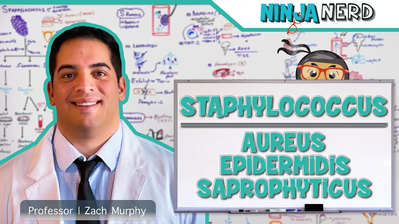
Staphylococcus: Aureus, Epidermidis, Saprophyticus

Short Summary:
This video discusses the three main species of Staphylococcus bacteria: aureus, epidermidis, and saprophyticus. It covers their identification (Gram-positive, catalase-positive, morphology), typical locations in the human body, pathogenic mechanisms (biofilms and exotoxins), the diseases they cause, and antibiotic treatment strategies, including the development of antibiotic resistance (MRSA, VRSA). Specific diagnostic tests like coagulase test, mannitol salt agar, urea broth, and novobiocin sensitivity are detailed. The video emphasizes the importance of understanding these bacteria's role in various infections, particularly those associated with medical devices.
Detailed Summary:
The video is structured as follows:
Section 1: Introduction and Staphylococcus Characteristics: The video begins with an engaging introduction, encouraging viewers to consult accompanying notes and illustrations on the presenter's website. It then defines Staphylococcus as a gram-positive, catalase-positive bacteria arranged in grape-like clusters. It highlights their non-motile and facultative anaerobic nature.
Section 2: Location and Types of Staphylococcus: Three species are detailed: S. aureus (common skin colonizer, especially in the nares), S. epidermidis (more prevalent skin flora than aureus), and S. saprophyticus (thrives in decaying organic matter, colonizes the perineum and female urinary tract). The presenter uses memorable phrases like "I'd rather get kicked in the perineal region" to help remember S. saprophyticus' location.
Section 3: Catalase and Coagulase Tests: The video explains the catalase test (hydrogen peroxide bubbling) as a way to identify Staphylococcus, noting that all three species are catalase-positive. The coagulase test (fibrinogen to fibrin conversion, causing clumping) is explained as a way to differentiate S. aureus (coagulase-positive) from the other two (coagulase-negative). The mannitol salt agar test, producing golden-yellow colonies for S. aureus, is also described. The term "golden yellow" is linked to the "aureus" in the species name.
Section 4: Differentiating S. epidermidis and S. saprophyticus: Both S. epidermidis and S. saprophyticus are catalase-positive and coagulase-negative. The urea broth test (urease enzyme producing a pink color) shows both are urease-positive. Novobiocin sensitivity differentiates them: S. epidermidis is sensitive, while S. saprophyticus is resistant.
Section 5: Pathogenic Mechanisms of S. aureus: The video details S. aureus' pathogenic mechanisms: biofilm formation (exopolysaccharide layer, leading to catheter-associated infections), and exotoxin production. Specific exotoxins are discussed: toxic shock syndrome toxin-1 (TSS-1, causing toxic shock syndrome with rash, hypotension, and fever), leukocidin (causing necrotizing pneumonia), exfoliative toxin (causing staphylococcal scalded skin syndrome or Ritter's disease), beta-hemolysin (destroying red blood cells), and enterotoxin (causing gastroenteritis). The presenter emphasizes the importance of understanding these toxins and their effects. A "positive Nikolsky's sign" is mentioned in relation to staphylococcal scalded skin syndrome.
Section 6: Pathogenic Mechanisms of S. epidermidis and S. saprophyticus: S. epidermidis primarily causes infections through biofilm formation on medical devices (catheters, prosthetic valves, joints). It's highlighted as a common contaminant in blood cultures. S. saprophyticus also forms biofilms, but its urease enzyme raises urine pH, promoting bacterial growth and potentially leading to struvite stone formation, causing urinary tract infections (UTIs).
Section 7: Diseases Caused by Staphylococcus Species: The video details the diseases caused by each species. S. aureus infections range from skin and soft tissue infections (furuncles, carbuncles, impetigo, cellulitis, abscesses) to more serious conditions like osteomyelitis, septic arthritis, bacteremia, septicemia, meningitis, brain abscesses, pneumonia, and infective endocarditis. The role of IV drug use and surgery in spreading S. aureus is emphasized. S. epidermidis causes catheter-associated infections, UTIs, prosthetic valve and joint infections. S. saprophyticus primarily causes UTIs (cystitis and pyelonephritis).
Section 8: Treatment and Antibiotic Resistance: The video discusses antibiotic resistance in Staphylococcus, focusing on S. aureus. It explains the roles of beta-lactamase, methicillin resistance (MRSA, due to the mecA gene and altered penicillin-binding protein 2a), and vancomycin resistance (VRSA, due to the vanA gene). Appropriate antibiotic choices for methicillin-sensitive S. aureus (MSSA), MRSA (hospital-acquired vs. community-acquired), and VRSA are detailed. Treatment for S. epidermidis infections often involves device removal, along with antibiotics (oxacillin, nafcillin, vancomycin). For S. saprophyticus UTIs, first-line antibiotics (nitrofurantoin, trimethoprim-sulfamethoxazole, fosfomycin) are discussed, with cephalexin, augmentin, and ciprofloxacin mentioned as second-line options. The video concludes with a reiteration of the key concepts and a closing remark.